Home
Uncategories
Back Bones Diagram / How The Spinal Cord Works Orthopedic Sports Medicine / Strong muscles and bones, flexible tendons and ligaments, and sensitive nerves contribute to a healthy spine.
Back Bones Diagram / How The Spinal Cord Works Orthopedic Sports Medicine / Strong muscles and bones, flexible tendons and ligaments, and sensitive nerves contribute to a healthy spine.
Back Bones Diagram / How The Spinal Cord Works Orthopedic Sports Medicine / Strong muscles and bones, flexible tendons and ligaments, and sensitive nerves contribute to a healthy spine.. Human skeleton with each bone name It is like that for several reasons, all of which you can understand by looking at the anatomy of the thoracic spine. Cervical bones diagram 12 photos of the cervical bones diagram cervical bones diagram, cervix anatomy. The fishbone diagram, also known as an ishikawa diagram, identifies possible causes for an effect or problem. The first seven bones (vertebrae) of your spine form your neck.
The vast difference in height and limb length between birth and adulthood are mainly the result of endochondral ossification in the. It consists of 5 lumbar vertebra that are numbered 1 through 5 from top to bottom i.e. At the back of each bone in the spine (vertebra) are bony points called processes, which muscles attach to. Our latest youtube film is ready to run. More commonly known as the shoulder blade, the scapula is a flat triangular bone located in the upper back.
Diagram Of A Human Spine Illustration Royalty Free Cliparts Vectors And Stock Illustration Image 95715300 from previews.123rf.com Anatomy of the spine southern california orthopedic institute. But, they are common in the back and can cause pain. Hip bones diagram of back and hip bones 9 out of 10 based on 30 ratings. For more anatomy content please follow us and visit our website: Spinal vertebrae bone spine vertebra toracica spinal cord spine structure back diagram spine sections spinal cord vertebrae spinal structure health diagram. The vast difference in height and limb length between birth and adulthood are mainly the result of endochondral ossification in the. Urinary system of the lower torso. Human back bones diagram poster 28 inch x 24 inch 16 inch x 13 inch.
Vertebrae separated by intervertebral discs.
Atlas (c1) the atlas is the first cervical vertebra and therefore abbreviated c1. Bone science human diagram anchor chart human body health back skeleton. Human back muscles and bones 12 photos of the human back muscles and bones human back muscles and bones, bone, human back muscles and bones. These bones are connected at the back with specialized joints. These bones work together to provide. This vertebra supports the skull. Spinal vertebrae bone spine vertebra toracica spinal cord spine structure back diagram spine sections spinal cord vertebrae spinal structure health diagram. The vertebral column houses the spinal canal, a cavity that. L1, l2, l3, l4, and l5. Spinal anatomy is a remarkable combination of strong bones, flexible ligaments and tendons, large muscles and highly sensitive nerves. The vertebral column is a series of approximately 33 bones called vertebrae, which are separated by intervertebral discs. Human back bones diagram poster 28 inch x 24 inch 16 inch x 13 inch. Muscle or tendon injuries can occur anywhere in the body.
Related posts of human back bones diagram female pelvis bones images. The vast difference in height and limb length between birth and adulthood are mainly the result of endochondral ossification in the. Hip bones diagram of back and hip bones 9 out of 10 based on 30 ratings. Posted on april 4, 2019. Back anatomy diagram lower bones rear view of human skeletal system showing upper back stock photo anatomy of the spine and back anatomy of the back bones sciences.
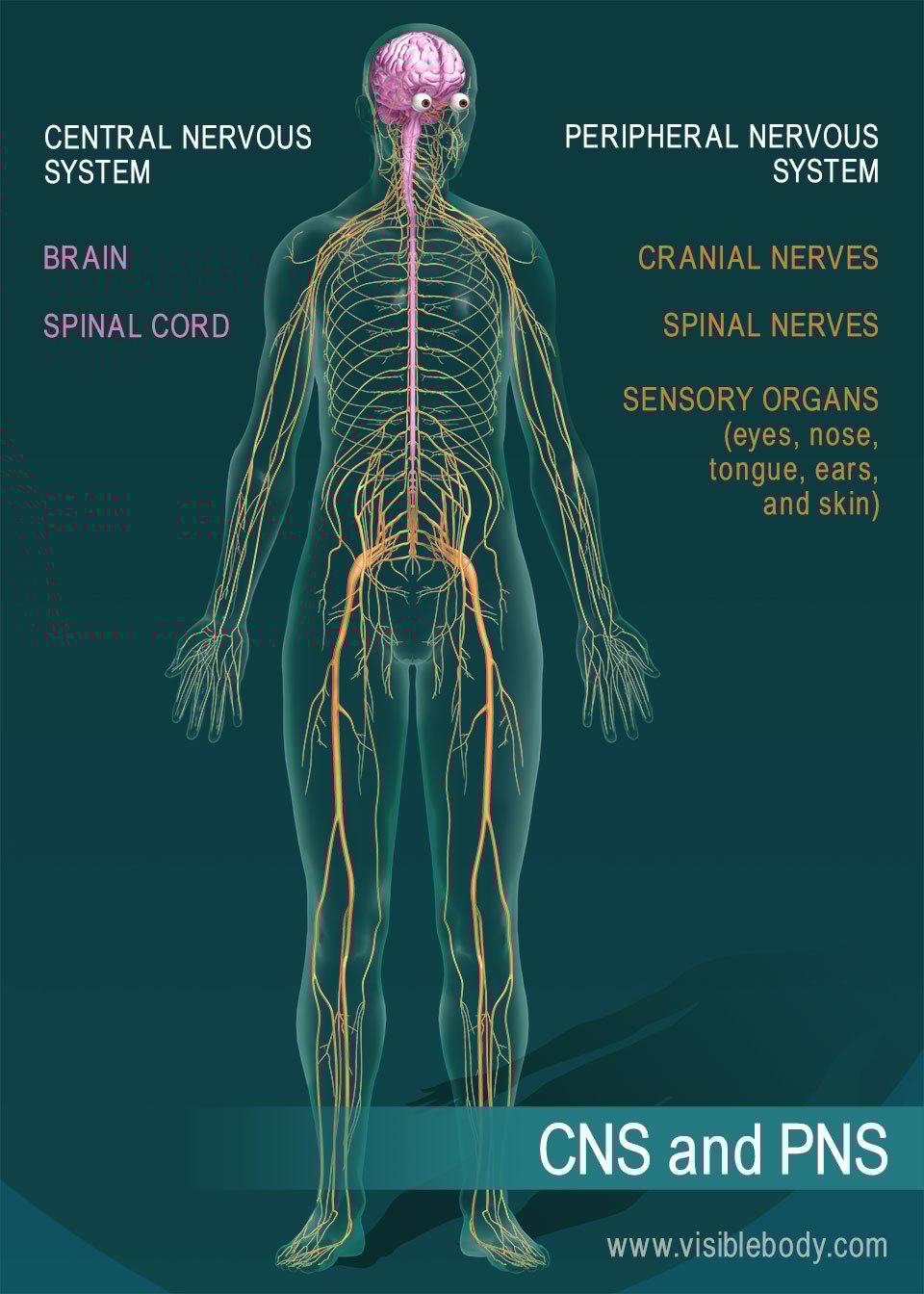
Nervous System Overview from www.visiblebody.com Indeed the practice of showing explicitness on the lower back has been performed for centuries. It connects with the collarbone at the front of the body. Vertebrae separated by intervertebral discs. Spinal anatomy is a remarkable combination of strong bones, flexible ligaments and tendons, large muscles and highly sensitive nerves. The cranial bones include occipital bone, two parietal bones, frontal bone, two temporal bones, sphenoid bone, and the ethmoid bone. We are pleased to provide you with the picture named anatomy of back muscles diagram.we hope this picture anatomy of back muscles diagram can help you study and research. Hip bones diagram of back and hip bones 9 out of 10 based on 30 ratings. The anatomy of the lumbar spine is quite complex.
The basics of back pain and spinal anatomy.
Atlas (c1) the atlas is the first cervical vertebra and therefore abbreviated c1. More commonly known as the shoulder blade, the scapula is a flat triangular bone located in the upper back. Anatomy of the spine southern california orthopedic institute. All the images are in vector format, allowing an optimal web display with zoom and shifting of the anatomical images. For more anatomy content please follow us and visit our website: The lumbar spine connects to the thoracic spine above and the hips below. Strong muscles and bones, flexible tendons and ligaments, and sensitive nerves contribute to a healthy spine. The atlas is the topmost vertebra, and along with c2, forms the joint connecting the skull and spine. The lumbar spine makes up the the lower end of the spinal column. Muscle or tendon injuries can occur anywhere in the body. It consists of 5 lumbar vertebra that are numbered 1 through 5 from top to bottom i.e. The atlas is a ring of bone made up of two lateral masses joined at. Lower jaw (mandible) collar bone.
Human back muscles and bones 12 photos of the human back muscles and bones human back muscles and bones, bone, human back muscles and bones. Human back bones diagram poster 28 inch x 24 inch 16 inch x 13 inch. The atlas is a ring of bone made up of two lateral masses joined at. Anatomical diagrams of the spine and back. The column can be divided into five different regions, with each region characterised by a different vertebral structure.
Lower Back Pain Causes Herniated Disc Bulging Disc from www.afcchiropractic.com Can you feel the bumps of your vertebrae along your back? The atlas is the topmost vertebra, and along with c2, forms the joint connecting the skull and spine. But, they are common in the back and can cause pain. This diagram depicts simple human body diagram. The radius is the bone which is present laterally, which mean. The spine diagram the spine diagram shown below, consists of many bones or vertebrae,soft discs,the spinal cord, and spinal nerves. L1, l2, l3, l4, and l5. We are pleased to provide you with the picture named anatomy of back muscles diagram.we hope this picture anatomy of back muscles diagram can help you study and research.
At the back of each bone in the spine (vertebra) are bony points called processes, which muscles attach to.
Nerves of the abdomen lower back and pelvis. Human back muscles and bones. Vertebrae there are 12 vertebrae in the thoracic spine. Strong muscles and bones, flexible tendons and ligaments, and sensitive nerves contribute to a healthy spine. It is like that for several reasons, all of which you can understand by looking at the anatomy of the thoracic spine. This spinal column provides the main support for your body, allowing you to stand upright, bend, and twist, while protecting the spinal cord from injury. The vast difference in height and limb length between birth and adulthood are mainly the result of endochondral ossification in the. This diagram depicts simple human body diagram. Bones of the pelvis and lower back. This vertebra supports the skull. The cranial bones include occipital bone, two parietal bones, frontal bone, two temporal bones, sphenoid bone, and the ethmoid bone. Human skeleton with each bone name Our latest youtube film is ready to run.


0 Comments:
Posting Komentar